
 American Heart Association
3 Days Only! Share Your Heart Challenge
Share your heart and give before midnight Friday to help unlock an extra $100,000 challenge gift!

November is American Diabetes Month

Need help managing your Type 2 diabetes? It’s a great time to learn about your risk for heart disease and stroke – and how to be an active member of your health care team.
Flu and RSV Prevention

It can be difficult to distinguish seasonal respiratory illnesses because they share many symptoms. Lower your chance of getting sick and protect your heart health.
Scientific Sessions 2023 – Register Today

Calling cardiovascular science and medicine professionals! Register now and join us for Scientific Sessions 2023 in Philadelphia, PA November 10-13. Network with colleagues, engage with experts and experience world class scientific discovery and practice changing educational content.
3 Days Only: Help unlock a $100,000 CHALLENGE GIFT!
Make your lifesaving gift before midnight Friday to help unlock a $100,000 challenge gift during the SHARE YOUR HEART challenge!

Healthy, Delicious Fall Recipes

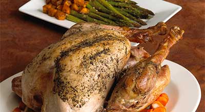
Roasted Turkey with Butternut Squash and Asparagus
While this may be the star of the Thanksgiving table, you don't have to wait until then to try this recipe!
View recipe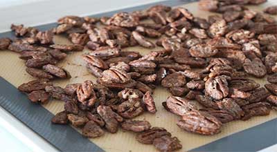
Maple-Spiced Pecans
Like other nuts, pecans are a good source of fiber, protein and antioxidants. This satisfying snack is sweet without the need for excess added sugar.
View recipe
Indian-Spiced Pumpkin Apple Soup
Garam masala adds warmth to this creamy soup that’s perfect for lunch on a chilly fall day.
View recipe
News and Stories
Helpful information about your health
从啊哈新闻阅读更多


Discover a Simpler Way to Give
The American Heart Association Donor Advised Fund Program offers an easy, flexible and tax-wise way to support all your favorite charities through one account.
Join us for a Heart Walk in your area
Heart Walk is the American Heart Association's premiere event for raising funds to help save lives from heart disease and stroke.